“Today’s approval is an important advance in cell therapy treatment in patients with blood cancers,” Peter Marks, M.D., Ph.D., director of the FDA Center for Biologics Evaluation and Research, said in a statement. “Hastening the return of the body’s white blood cells can reduce the possibility of serious or overwhelming infection associated with stem cell transplantation.”
New research out of the University of Cincinnati examines the impact that maternal stress during pregnancy has on the neurodevelopment of babies.
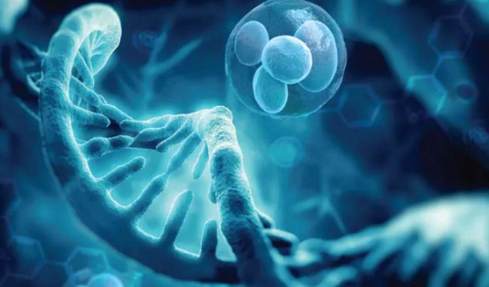
Prenatal maternal stress life events are associated with adverse neurodevelopmental outcomes in offspring. Biological mechanisms underlying these associations are largely unknown, but a chemical reaction in the body in which a small molecule known as a methyl group gets added to DNA, called DNA methylation, likely plays a role, according to researchers. These findings could provide new insights into how the fetal environment potentially influences not only neurodevelopment, but metabolism and immunologic functions as well.
More than 5,500 people took part in the study with that population broken down into 12 separate cohorts, according to Anna Ruehlmann, a postdoctoral fellow in the Department of Environmental and Public Health Sciences in the UC College of Medicine and lead author of the research.
As part of an immune response, white blood cells release web-like Neutrophil Extracellular Traps (NETs) to capture and kill pathogens. While typically beneficial, Yost had previously shown that overactive NETs exacerbate certain illnesses. In conditions such as overwhelming infection, NETs can clog blood vessels and lead to inflammatory tissue damage.
To determine whether NETs could be responsible for complications seen in COVID-19, the team examined plasma from 33 patients, along with tracheal aspirates from the lungs. They found that NET activity correlated with disease severity.
Patients on life support and those who died from COVID-19 had significantly more signs of NET activation than patients who were not as sick or who went on to recover. The NET immune response was lower still in healthy people. NET levels also tracked with a marker for blood-oxygen levels, an independent indicator of disease severity.
Similarly, plasma from sick patients was primed to launch the NET response. When examined in laboratory experiments, plasma from COVID-19 patients triggered white blood cells from healthy patients to shoot out 50 times as many NETs as cells exposed to plasma from otherwise healthy adults.
“This study may tell us that NET levels in the blood could potentially help predict disease severity and mortality in COVID-19,” says Yost, a physician-scientist at U of U Health. “Additional information is urgently needed in this pandemic regarding how to know which patient will fare better or worse.” Larger studies will need to be done to determine whether NETs could become a biomarker for COVID-19 severity. “Importantly, we think exaggerated NETs could be a cause of morbidity and mortality in COVID-19,” Yost says.
In support of the idea, collaborators at Cold Spring Harbor Laboratory showed that blood vessels in the lungs of deceased COVID-19 patients were dotted with clumps of NET-producing cells and a critical type of blood cell for clotting, the platelets. Another recent study from U of U Health showed that platelets become hyperactive during the disease. Investigations are now underway to determine whether NETs and platelets increase the risk for blood clotting and other clinical manifestations of COVID-19.
There they met Dr. Joe Rossano, who heads up the cardiac center at CHOP.
“Yeah, they’re a very complicated surgery,” Dr. Rossano says. “You know, prior to the 1980s and ’90s, there were essentially no good surgical options for these patients. But a number of very innovative surgical techniques were developed that have allowed many of these children to survive and thrive.”
Children born with HLHS go through a series of three surgeries to essentially rework the plumbing of the heart. The first surgery comes within days of birth. The second surgery generally occurs three to six months later. And the third surgery is usually performed about three years later.
This series of surgeries allows children with HLHS to live relatively normal lives, but it isn’t perfect.
Having the right side of the heart perform the tasks normally handled by both sides of the heart puts tremendous stress and pressure on the right side.
As patients with HLHS are aging, doctors are realizing many of their hearts are unable to continue to function on their own, and some patients need a heart transplant.
But Andrea Sexton found out about a clinical trial going on at Mayo Clinic aiming to solve that problem.
“Quickly they set me up with an interview over the phone with Dr. Nelson,” Andrea Sexton says. “He was amazing. He gave me hope.”
Dr. Tim Nelson oversees the Todd and Karen Wanek Family Program for Hypoplastic Left Heart Syndrome. He leads a team of roughly 60 people who research and conduct clinical trials for new HLHS treatments.
“The HLHS program has a mission to … recreate the right ventricle to make it bigger and stronger,” Dr. Nelson says. “So we’re finding ways of inventing new therapies to make that right heart stronger by stimulating the growth of the heart muscle and make the five-horsepower engine, a 10-horsepower engine, a 50-horsepower engine. And if we make it strong enough, we believe that that has a shot at delaying and preventing transplant for a significant number of these children.”
The key to this groundbreaking regenerative therapy clinical trial is stem cells, but not just any stem cells. Dr. Nelson’s team uses the babies’ own stem cells derived from umbilical cord blood.
“So the first product that we’re testing in our clinical trial is using umbilical cord blood from the baby’s own body,” Dr. Nelson says. “So the child has to be diagnosed in utero, and we have to be able to collect the cord blood at birth. We can collect the cord blood from anywhere in the country and ship it into Rochester, Minnesota, to have it processed to have a high concentrated product that gets frozen in low temperatures of liquid nitrogen. That product is frozen for three months until the child has their second surgery, or the Glenn operation, which then we can bring those cells back into the operating room, thaw them, and deliver them directly into the heart muscle.”
“The hope of this is that it causes a fertilizer type of an effect where these cells are able to fertilize the right ventricle muscle and allow it to grow bigger and stronger because it’s received this cell-based therapy,” Dr. Nelson says.
On May 4, 2017, Ryals became the fifth clinical trial patient in Mayo Clinic’s HLHS team’s research.
Dr. Nelson and his team, who had traveled to Philadelphia to deliver the cells, looked on as a surgeon from CHOP injected the cells into Ryals’ tiny heart near the end of his operation. A surgical assistant counted down as the surgeon injected the cells into different areas of the heart.
After several tense hours of waiting during Ryals’ surgery, his parents were emotional and relieved to hear all went well and that there were no problems injecting Ryals’ cells.
During an emotional hug, tears flowed down Andrea Sexton’s face as she thanked Dr. Nelson.
“You’re welcome,” Dr. Nelson says. “He’s a trooper.”